
Adaptive structures proliferated in the rabbit shoulder after 8 weeks from the insertion of a titanium implant
All claims expressed in this article are solely those of the authors and do not necessarily represent those of their affiliated organizations, or those of the publisher, the editors and the reviewers. Any product that may be evaluated in this article or claim that may be made by its manufacturer is not guaranteed or endorsed by the publisher.
Accepted: 23 February 2022
Authors
Aim A special place in the research related to implantology is held by those who study the process of osseointegration of implants. The proper development of the osseointegration process is crucial for the survival rate of the implant. The chances of long-term survival are related to the structure of the bone in which the implant is inserted.
Materials and methods In three 10-month-old male rabbits, self-tapping titanium implants with a diameter of 2 mm were inserted in the femoral shaft. After 8 weeks, the rabbits were stuk and the fragment of the femur containing the implant was collected. The collected pieces were fixed in Stieve mixture, embedded in paraffin, and the 5 µm sections obtained were stained with the Goldner trichrome method.
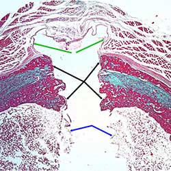
Results The microscopic examination revealed that the bone proliferated around the implant had two starting points, the periosteum and the endosteum. The newly proliferated bone from the periosteal level gradually extends to the middle area of the interface, outward over a certain distance and subperiosteal over another distance. The proliferated bone at the endosteal level extends to the middle area of the interface, towards the medullary canal to a certain depth, but also subendosteal to a certain distance. Moreover, the newly proliferated bone shows a clear tendency to reshape into Haversian bone, at a clearly higher level above the existing structure in the bone distant from the implantation area. This aspect shows the tendency of a bone with greater strength than the rest of the bone to form around the implant.
Conclusions The increase of the contact surface at the bone-implant interface as well as the proliferation of a bone with high resistance are the result of an adaptive reaction to restore the resistance of the area, weakened following the trauma caused by the insertion of the implant.
Downloads
Citations
How to Cite

This work is licensed under a Creative Commons Attribution-NonCommercial 4.0 International License.