Evaluation of color stabilization of peri-implant mucosa in different restorative material, abutment and tissue thickness combinations: an in vitro study
All claims expressed in this article are solely those of the authors and do not necessarily represent those of their affiliated organizations, or those of the publisher, the editors and the reviewers. Any product that may be evaluated in this article or claim that may be made by its manufacturer is not guaranteed or endorsed by the publisher.
Authors
Aim The aim of this in vitro study is to determine the level of color change of the peri-implant mucosa in several combinations of parameters, including different experimentally-created peri-implant tissue thicknesses, and different prosthetic crown and abutment materials, by spectrophotometric measurements.
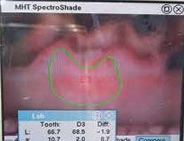
Materials and methods In this in vitro study, a sheep’s head was used because it resembles human mucosa in terms of color and texture. Different experimentally-created peri-implant tissue thicknesses were determined, i.e. 1, 1.5, 2 and 3 mm, and to provide these thicknesses 0.5, 1 and 2 mm thick connective tissue grafts were harvested from the palatal mucosa of the sheep’s head. These grafts were placed under the mucosal flap and fixed with tissue adhesive. Titanium and zirconia were chosen as abutment materials. Metal-porcelain crowns, zircon crowns and feldspathic porcelain crowns were selected as crown materials. Materials were represented by 5 x 5 x 1 mm blocks made of the same materials. For each study group, two measurements were made using a spectrophotometer. The first measurement determined the color of the flap in different experimentally-created tissue thicknesses, and the second measurement determined the color of the tissue after the prosthetic material and abutment material were placed under these flaps. Statistical comparison of the two measurement values was used to determine the color change.
Results Spectrophotometer measurements show that the naked eye could distinguish between all groups when the mucosa thicknesses were 1 and 1.5 mm. When the mucosa thickness was 2 mm, color change was observed in the titanium abutment and prosthesis groups, while color stability was achieved in the zircon abutment and prosthesis groups when the mucosa thickness was 3 mm.
Conclusions Within the limits of this study, peri-implant mucosa thickness is an important factor in color stabilisation; mucosa thickness must be a minimum of 2 mm to achieve this stabilisation.
Downloads
Citations
How to Cite

This work is licensed under a Creative Commons Attribution-NonCommercial 4.0 International License.